
When people search for the word diag image, they often mean two things. Some use it for diagnostic imaging, like X-rays, CT scans, or MRI scans that doctors use to look inside the body. Others use it for a diagrammatic image, which is a simple visual drawing that explains an idea, process, or system. Both meanings share one purpose: making things clearer.
In healthcare, a diag image can save lives. Doctors rely on these scans to detect problems early, guide treatments, and monitor recovery. Without them, many conditions would stay hidden until it is too late.
In schools and workplaces, diagrammatic images play a different but still powerful role. A good diagram can explain in seconds what pages of text may struggle to show. Complex systems, business plans, or even science lessons become easier to understand when broken down into a visual format.
This article takes a close look at both sides of the word diag image. It explains what diagnostic imaging means, the different methods doctors use, and the benefits and risks. It also explores diagrammatic images, why they are important in learning and communication, and how both worlds now come together with new technology.
By the end, you will have a clear picture of why diag images matter in health, education, and daily life, and how they are shaping the way we see and understand the world.
Two Interpretations oOf “Diag Image”
The word diag image has two main meanings. Both are important, but they are used in very different ways.
Interpretation A: Diagnostic Imaging (Medical Context)
In medicine, a diag image usually means diagnostic imaging. These are pictures of the inside of the body taken by machines like:
- X-ray for bones and chest scans
- CT scans for cross-section views of organs
- MRI scans for soft tissues like the brain or muscles
- Ultrasound for real-time pictures using sound waves
- PET scans for checking body functions with special tracers
Doctors use these images to diagnose diseases, monitor ongoing conditions, and plan treatments. A broken bone, a heart problem, or even a hidden tumor can often be found with a diag image before symptoms become too serious.
Interpretation B: Diagrammatic Image (Educational and Technical Context)
Outside of medicine, a diag image can also mean a diagrammatic image. These are visuals created to explain ideas or systems. Common examples include:
- Flowcharts for step-by-step processes
- Concept maps for linking ideas together
- System diagrams for engineering or software design
- Infographics for turning data into visuals
- Explanatory visuals for simplifying complex topics
The purpose of a diagrammatic image is simple: to make information easier to understand. A clear diagram can save time, reduce confusion, and help people learn faster. Whether in classrooms, business meetings, or technical manuals, a diag image turns difficult ideas into visuals that everyone can follow.
Diagnostic Imaging: Methods, Modalities, And Technology
A diag image in medicine is created through different imaging methods. Each method has its own role in showing what is happening inside the body.
X-Ray And Plain Radiography
X-rays are one of the oldest and most common imaging tools. They are quick, simple, and best for looking at bones, chest, and joints. A broken bone or a chest infection is often detected with a plain X-ray.
Computed Tomography (CT)
A CT scan takes many X-ray images and combines them into cross-section “slices.” This gives doctors a more detailed picture of organs, blood vessels, and soft tissues. CT is useful for spotting tumors, injuries, or internal bleeding.
Magnetic Resonance Imaging (MRI)
MRI uses strong magnets and radio waves instead of radiation. It produces clear pictures of soft tissues such as the brain, spine, and muscles. This makes it safer for repeat scans and very useful for detecting nerve or brain problems.
Ultrasound / Sonography
Ultrasound uses sound waves to create real-time images. It is safe, non-invasive, and widely used in pregnancy, heart scans, and checking soft organs. Doctors can see movement and blood flow, which makes it helpful in many situations.
Nuclear Medicine And PET Scans
Nuclear imaging uses small amounts of radioactive tracers to show how organs and tissues are working. A PET scan can reveal early signs of cancer, heart disease, or brain disorders. Unlike other scans, it focuses more on function than structure.
Emerging Enhancements
Diagnostic imaging continues to advance. Today, hospitals use:
- 3D reconstructions to give a full picture of organs or bones.
- Volumetric imaging to measure size and shape more precisely.
- Multiplanar views so doctors can look at scans from different angles.
- AI-assisted tools that help radiologists spot details faster and reduce errors.
- Autonomous imaging systems and real-time computing, which improve speed and accuracy.
These improvements are making diag images more reliable, easier to interpret, and more useful in both diagnosis and treatment planning.
Diagnostic Imaging: Benefits And Challenges
A diag image is one of the most powerful tools in modern medicine. It offers many benefits but also brings certain challenges that doctors and patients need to keep in mind.
Benefits Of Diagnostic Imaging
- Non-invasive look inside the body – Scans like X-ray, CT, and MRI allow doctors to see internal organs and bones without surgery. This reduces risk, pain, and recovery time.
- Early detection and monitoring – Many diseases, such as cancer or heart problems, can be detected at an early stage through imaging. Doctors also use scans to monitor how treatments are working and adjust plans if needed.
- Improved accuracy with AI support – New AI-assisted tools highlight suspicious areas, speed up analysis, and reduce human error. This leads to quicker results and better patient outcomes.
Challenges Of Diagnostic Imaging
- Radiation exposure and safety – CT scans and X-rays use ionizing radiation. While generally safe, repeated scans can pose risks. Some tests also need contrast agents, which may not be suitable for all patients.
- Interpretation and over-imaging – Not all findings are clear. Sometimes images show harmless changes, called “incidental findings,” that may cause stress or confusion. Overuse of imaging can also lead to unnecessary costs and procedures.
- Data management and privacy – Hospitals must store and share large amounts of imaging data. Systems like DICOM and PACS help, but issues with compatibility, speed, and patient privacy remain a challenge.
Overall, diag images are essential in modern healthcare. They give doctors strong insight into the body, but careful use, skilled interpretation, and safe data handling are just as important.
Diagrammatic Images: Design, Use Cases, And Tools
A diag image can also mean a diagrammatic image. These are simple visuals that explain ideas in a way words alone cannot. They break down complex topics, help people learn faster, and make communication clear.
Why Diagrammatic Images Are Powerful
- They simplify complexity. A well-made diagram shows structure or process at a glance.
- They support visual learners who remember images better than text.
- They bridge gaps between experts and non-experts. A diagram can explain technical details in plain visuals.
Common Use Cases
- Education – Teachers use diagrams in textbooks, online courses, and science lessons. A biology diagram or an engineering chart makes hard subjects easier.
- Business and Presentations – Flowcharts, org charts, and project timelines guide teams and improve planning.
- Scientific and Technical Communication – Engineers and software developers use system diagrams or process flows to show how things work.
- Healthcare Communication – Diagrams of the human body or treatment steps help patients understand medical advice.
Tools And Technologies For Making Diag Images
- Traditional tools – Whiteboards, pen and paper are still effective for brainstorming and quick sketches.
- Digital tools – Software like Canva, Lucidchart, Microsoft Visio, and Figma makes it easy to design professional visuals.
- AI-assisted tools – New platforms can auto-layout diagrams, suggest schematics, or even turn text into diagrams. This saves time and ensures clarity.
A diag image in this form is more than just a drawing. It is a bridge between knowledge and understanding, making information easier to share and remember.
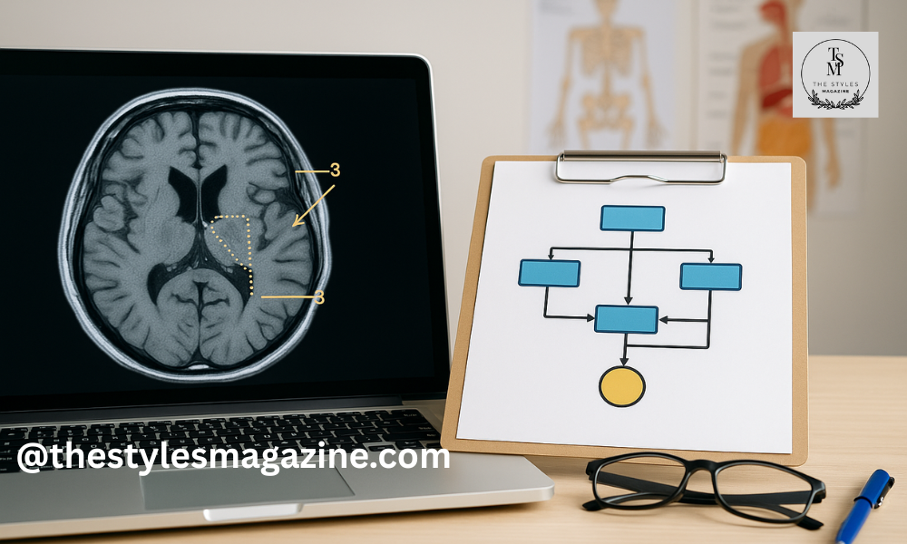
Bringing Them Together: Hybrid “Diag Image” Applications
In many cases, a diag image is not only a scan or a diagram but a mix of both. By combining medical imaging with diagrammatic visuals, doctors and educators can explain complex information in ways that are easier to understand.
When Medical Images Are Combined With Diagrams
- Annotated scans – An MRI or CT image can be marked with arrows, labels, or colored regions to point out key details.
- Educational visuals – Students learn better when a real diagnostic image is paired with a clear diagram. For example, a brain MRI next to a labeled sketch helps connect theory to reality.
- Patient reports – Radiologists often add simple diagrammatic reconstructions to explain results in a way patients can follow without medical training.
Technology Trends Driving Hybrid Use
- 3D reconstructions and multiplanar views make it possible to rotate, slice, and explore the body from every angle.
- AI and computer-aided diagnosis (CAD) systems can highlight suspicious areas, generate labels, and create simplified overlays that non-specialists can understand.
- Cloud-based viewers like Weasis and web annotation platforms allow real-time sharing, teaching, and collaboration across locations.
This hybrid approach makes diag images more than just pictures. They become teaching tools, decision aids, and communication bridges between experts and everyday people.
Best Practices And Tips For Creating or Interpreting A “Diag Image”
A diag image is most useful when it is designed with care and interpreted with skill. Whether you are creating a diagrammatic image for learning or analyzing a medical scan, following best practices ensures accuracy, clarity, and trust.
Designing A Diagrammatic Diag Image
- Know your audience – Decide if the image is for experts or beginners. A diagram for doctors can include medical terms, while one for patients should use simpler labels.
- Balance accuracy with simplicity – Show only what matters. Avoid clutter, highlight the key elements, and keep labels clear.
- Stay consistent – Use the same colors, shapes, and symbols across diagrams. Consistency makes information easier to follow.
- Check readability – Make sure the image works on small screens, large screens, or printed pages.
- Be ethical with medical visuals – If you combine diagnostic scans with diagrams, credit the source, annotate carefully, and always protect patient privacy.
Interpreting A Medical Or Hybrid Diag Image
- Start with the raw scan – Look at the original image before relying on overlays or labels.
- Know what is simplified – Some details may be left out in diagrammatic reconstructions. Understand what remains accurate and what is abstract.
- Cross-check reports – Use both the schematic overlays and the radiologist’s written notes for context.
- Stay cautious with AI tools – Automated labels or highlights are helpful but not final. They suggest possibilities, not diagnoses.
By designing and interpreting diag images with these practices in mind, professionals and learners can gain accurate insights without confusion, while maintaining trust and clarity.
Future Trends In “Diag Image” Technology
The world of diag images is changing fast. New tools are making both diagnostic scans and diagrammatic visuals more powerful and easier to use.
- AI-driven annotation – Artificial intelligence can now mark scans automatically, add labels, and even turn text into images or diagrams. This saves time and supports faster decisions.
- AR and VR integration – Augmented reality and virtual reality bring a new level of depth. Imagine a doctor looking at a live ultrasound with a virtual guide overlaid in real time.
- 3D and immersive imaging – From 3D biomedical visuals to interactive volumetric diagrams, diag images are moving beyond flat pictures. Users can rotate, zoom, and explore in detail.
- Cloud-based collaboration – Hospitals, schools, and research centers are using online platforms to share and annotate images together, making remote diagnostics and learning more effective.
- Democratization of imaging – Tools are no longer limited to experts. Patients, students, and non-specialists can now create and share diag images with easy apps, citizen science projects, and remote education platforms.
These trends point to a future where diag images are more accessible, interactive, and central to how we learn, teach, and treat.
Conclusion
The term diag image carries two clear meanings: diagnostic imaging in medicine and diagrammatic images in education and communication. While different, both serve the same purpose—turning complexity into clarity.
In healthcare, diag images give doctors a safe way to look inside the body, detect problems early, and guide treatments. In classrooms, businesses, and labs, diagrammatic visuals make information simple and easy to understand. When these two forms come together, they create hybrid solutions that support both professionals and everyday people.
As technology grows—through AI, cloud tools, 3D visuals, and immersive platforms—the role of diag images will continue to expand. They are no longer just pictures; they are tools for understanding, decision-making, and collaboration.
FAQs
What Does “Diag Image” Mean?
The term diag image can mean either a diagnostic medical image like an X-ray or MRI, or a diagrammatic image such as a chart or flowchart.
Why Are Diag Images Important In Healthcare?
Diag images help doctors see inside the body without surgery, detect diseases early, and plan the right treatments.
How Are Diag Images Used In Education?
In schools and training, diagrammatic diag images simplify complex ideas with visuals like charts, maps, or system diagrams.
Are Diag Images Safe For Patients?
Most diagnostic imaging methods are safe. Some use low radiation, like X-rays or CT scans, but doctors balance the risks with the benefits.
What Is The Future Of Diag Images?
AI, 3D visuals, and cloud tools are making diag images more accurate, interactive, and accessible for both experts and non-experts.
Thank you for exploring our Blog! For additional captivating content, feel free to explore the corresponding category.
Disclaimer: This article about diag image is for general information only. It should not be taken as medical or professional advice. For health concerns, always consult a qualified doctor. For technical or educational uses, seek expert guidance when needed.
Edivawer Review: How It’s Changing Education And Workplace Training